
By Danny Chan
The Morita Group is, without exaggeration, a vaunted institution in the global arena of X-ray diagnostics and endodontics. The company’s premium portfolio covers most major equipment you would find inside of a modern dental practice: Imaging systems; cone beam computed tomography; treatment units; laser and turbines; as well as endodontic measuring and preparation systems.
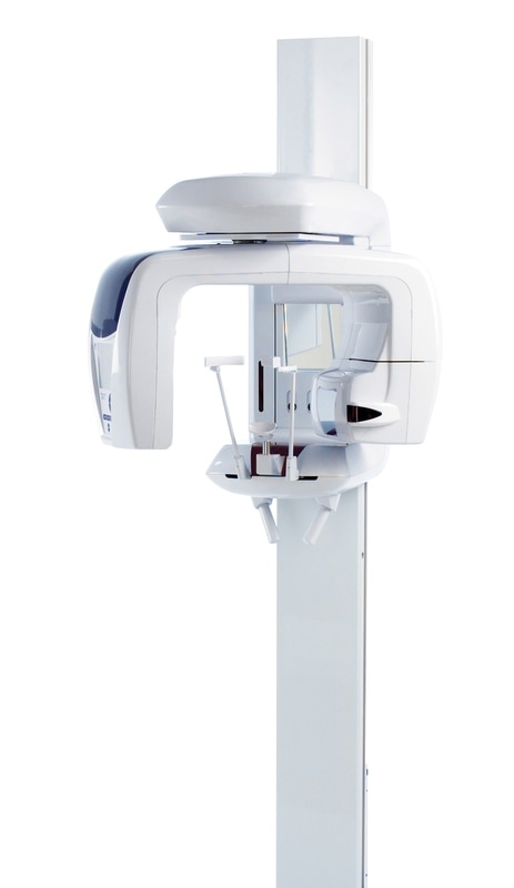
It has been 100 years since the Japanese dental manufacturer first embarked on its milestones-paved journey to produce high quality precision ware for an appreciative dental community. It is perhaps fitting to mark its centennial year, by celebrating one of Morita’s path-breaking innovations, one that that has in recent years been widely praised for pushing the frontiers of 3D CBCT technology – the Veraviewepocs 3D R100.
Veraviewepocs 3D R100 represents a bold new leap in the 3D world of x-ray diagnostics. Offering eight field of view options and Morita's world-renowned image quality, the CBCT unit is suitable for a wide variety of dental applications including implant planning. It features the groundbreaking 3D Reuleaux Full Arch fields of view (FOVs) that provide a unique shape for full arch imaging. More closely matching the natural dental arch form, the 3D Reuleaux allows a complete scan of the maxilla and/or the mandible while significantly reducing dosage by excluding areas outside the region of interest.
Speaking to dentists fortunate enough to own the Veraviewepocs 3D R100, a recurring sentiment is palpable: “I don’t know how I ever managed to work without it.” As 3D dental imaging deepens its roots, Dr David Cable, Specialist Endodontist, Dr David H Cable Endodontist, unabashedly compares its future to Apple’s ubiquitous mobile phone, asking earnestly:
“3D technology has so radically improved endodontic diagnosis that it is truly a paradigm shift.
It definitely equates with and, in my humble opinion, even surpasses the benefits we saw when microscopes were adopted into endodontics.”
“I would predict that every endodontic practice in Australia will own a CBCT unit within the next 5 years, just as happened with microscopes. It's just like your mobile phone. Can you imagine being without an iPhone these days?”
An early adopter of CBCT technology – David says his was the first specialist endodontic practice in Australia to install a CBCT unit at the start of 2013 – the tech-savvy endodontist spent two years “investigating the evolution”, testing various brands of the then nascent innovation. Prior to 2013, David’s practice had outsourced all 3D imaging to radiology practices.
“Our early cases were mainly imaged with medical MSCT at 1000µm resolution and doses of about 1-2000µSv. Accordingly, these were very selectively utilised for only a few special cases. Early dental CBCT units like iCat, with its typical 300µm to at best 200µm resolutions, had doses of nearly 200-300µSv. The image quality of these scans was limited and only helpful for a small number of patients.”
Recognising the move from MSCT to 3D-CBCT a decided leap, David’s first brush with the Morita unit was no less eye opening:
“The Veraviewepocs’ ability to image just a small 4 x 4cm FOV at 125µm at doses of 20-60µSv, results in a near 1000x improvement over MSCT and a 100x reduction in dose compared with medical imaging.”
“Most other CBCT units prior to Morita's 3De could only do whole head (23 x 17 cm) or double jaw (16 x 8 cm) scans. Most had 20-40 second scan times for so called "high resolution" modes but such long exposure times not only increased radiation dosage but also degraded the image quality due to motion artefact.”
By comparison, Morita unit’s scan time is only 9.4 seconds for a high-resolution scan, he proudly asserts.
“Some manufacturers say they have a short scan time but these are actually for low resolution or scout scans. Even units capable of high resolution produced way poorer scans, due to the increased time available for patient movement.”
Another convinced Veraviewepocs 3D R100 customer is Dr Stephen Chen, Periodontist, Melbourne Periodontics. The President-Elect of the International Team for Implantology (ITI) attributes the “excellent quality” of the 3D images to the fine pixel size:
“The fine pixel size and ability to select the field of view (FOV) offer significant advantages. For patients requiring single tooth implants or implants in small edentulous gaps, limiting the FOV has a positive advantage of reducing the radiation dose.”
“In relation to implant diagnosis, the R100 provides accurate and clear cross-sectional images for implant planning. The DICOM files are easily exported to proprietary 3-D software planning packages as well.”
The versatility of the x-ray unit allows it to be used for all dental implant assessment applications, Stephen qualifies.
“The most significant advantage for us is the ability to use a small field of view of approximately 40mm x 40mm. Most of our implant patients require single tooth implants or implants to replace short edentulous spans. The R100 is perfect for these case types.”
For David, differential pain diagnosis used to be a common – and often problematic – part of specialist endodontic practice, but now can be effectively solved with the aid of 3D imaging.
“Since our past appreciation of anatomy was limited to 2D imaging, we had to imagine in our minds what the anatomy of a tooth, a root, a canal or the surrounding anatomy may be.
Now we can actually "see" it all in 3D.”
Besides enhanced diagnostics, the dentists shared other Veraviewepocs’ practice-building benefits. Stephen says the ability to accurately assess a potential dental implant site and provide this information in a timely fashion has been the most significant change.
"Having the CBCT unit in the office has improved efficiency and workflow. It is now possible to provide the patient with a comprehensive assessment of the underlying bone and the visibility of placing implants during the preliminary consultation.”
“The move to a fully digital workflow for both conventional restorative dentistry as well as implant dentistry will be the key change in the paradigm for treatment care.”
For David, 3D imaging helped his specialist clinic ride with changes:
“We have seen a shift in the type of cases being referred to our practice. Sadly we get fewer primary RCT cases and we are getting more re-treatment and more difficult cases – resorption and pain differential diagnoses. 3D imaging has allowed us to keep pace with these changes and meet the increased challenges these pose.”
“Dentistry is shifting into a new era with ever increasing availability and reliance on technology. Twenty years ago, the only things improving were dental materials. Modern practices are now critically dependent upon technology and most simply cannot function without them. 3D imaging is definitely another part of this evolution.”
The Morita Group is, without exaggeration, a vaunted institution in the global arena of X-ray diagnostics and endodontics. The company’s premium portfolio covers most major equipment you would find inside of a modern dental practice: Imaging systems; cone beam computed tomography; treatment units; laser and turbines; as well as endodontic measuring and preparation systems.
It has been 100 years since the Japanese dental manufacturer first embarked on its milestones-paved journey to produce high quality precision ware for an appreciative dental community. It is perhaps fitting to mark its centennial year, by celebrating one of Morita’s path-breaking innovations, one that that has in recent years been widely praised for pushing the frontiers of 3D CBCT technology – the Veraviewepocs 3D R100.
Veraviewepocs 3D R100 represents a bold new leap in the 3D world of x-ray diagnostics. Offering eight field of view options and Morita's world-renowned image quality, the CBCT unit is suitable for a wide variety of dental applications including implant planning. It features the groundbreaking 3D Reuleaux Full Arch fields of view (FOVs) that provide a unique shape for full arch imaging. More closely matching the natural dental arch form, the 3D Reuleaux allows a complete scan of the maxilla and/or the mandible while significantly reducing dosage by excluding areas outside the region of interest.
Speaking to dentists fortunate enough to own the Veraviewepocs 3D R100, a recurring sentiment is palpable: “I don’t know how I ever managed to work without it.” As 3D dental imaging deepens its roots, Dr David Cable, Specialist Endodontist, Dr David H Cable Endodontist, unabashedly compares its future to Apple’s ubiquitous mobile phone, asking earnestly:
“3D technology has so radically improved endodontic diagnosis that it is truly a paradigm shift.
It definitely equates with and, in my humble opinion, even surpasses the benefits we saw when microscopes were adopted into endodontics.”
“I would predict that every endodontic practice in Australia will own a CBCT unit within the next 5 years, just as happened with microscopes. It's just like your mobile phone. Can you imagine being without an iPhone these days?”
An early adopter of CBCT technology – David says his was the first specialist endodontic practice in Australia to install a CBCT unit at the start of 2013 – the tech-savvy endodontist spent two years “investigating the evolution”, testing various brands of the then nascent innovation. Prior to 2013, David’s practice had outsourced all 3D imaging to radiology practices.
“Our early cases were mainly imaged with medical MSCT at 1000µm resolution and doses of about 1-2000µSv. Accordingly, these were very selectively utilised for only a few special cases. Early dental CBCT units like iCat, with its typical 300µm to at best 200µm resolutions, had doses of nearly 200-300µSv. The image quality of these scans was limited and only helpful for a small number of patients.”
Recognising the move from MSCT to 3D-CBCT a decided leap, David’s first brush with the Morita unit was no less eye opening:
“The Veraviewepocs’ ability to image just a small 4 x 4cm FOV at 125µm at doses of 20-60µSv, results in a near 1000x improvement over MSCT and a 100x reduction in dose compared with medical imaging.”
“Most other CBCT units prior to Morita's 3De could only do whole head (23 x 17 cm) or double jaw (16 x 8 cm) scans. Most had 20-40 second scan times for so called "high resolution" modes but such long exposure times not only increased radiation dosage but also degraded the image quality due to motion artefact.”
By comparison, Morita unit’s scan time is only 9.4 seconds for a high-resolution scan, he proudly asserts.
“Some manufacturers say they have a short scan time but these are actually for low resolution or scout scans. Even units capable of high resolution produced way poorer scans, due to the increased time available for patient movement.”
Another convinced Veraviewepocs 3D R100 customer is Dr Stephen Chen, Periodontist, Melbourne Periodontics. The President-Elect of the International Team for Implantology (ITI) attributes the “excellent quality” of the 3D images to the fine pixel size:
“The fine pixel size and ability to select the field of view (FOV) offer significant advantages. For patients requiring single tooth implants or implants in small edentulous gaps, limiting the FOV has a positive advantage of reducing the radiation dose.”
“In relation to implant diagnosis, the R100 provides accurate and clear cross-sectional images for implant planning. The DICOM files are easily exported to proprietary 3-D software planning packages as well.”
The versatility of the x-ray unit allows it to be used for all dental implant assessment applications, Stephen qualifies.
“The most significant advantage for us is the ability to use a small field of view of approximately 40mm x 40mm. Most of our implant patients require single tooth implants or implants to replace short edentulous spans. The R100 is perfect for these case types.”
For David, differential pain diagnosis used to be a common – and often problematic – part of specialist endodontic practice, but now can be effectively solved with the aid of 3D imaging.
“Since our past appreciation of anatomy was limited to 2D imaging, we had to imagine in our minds what the anatomy of a tooth, a root, a canal or the surrounding anatomy may be.
Now we can actually "see" it all in 3D.”
Besides enhanced diagnostics, the dentists shared other Veraviewepocs’ practice-building benefits. Stephen says the ability to accurately assess a potential dental implant site and provide this information in a timely fashion has been the most significant change.
"Having the CBCT unit in the office has improved efficiency and workflow. It is now possible to provide the patient with a comprehensive assessment of the underlying bone and the visibility of placing implants during the preliminary consultation.”
“The move to a fully digital workflow for both conventional restorative dentistry as well as implant dentistry will be the key change in the paradigm for treatment care.”
For David, 3D imaging helped his specialist clinic ride with changes:
“We have seen a shift in the type of cases being referred to our practice. Sadly we get fewer primary RCT cases and we are getting more re-treatment and more difficult cases – resorption and pain differential diagnoses. 3D imaging has allowed us to keep pace with these changes and meet the increased challenges these pose.”
“Dentistry is shifting into a new era with ever increasing availability and reliance on technology. Twenty years ago, the only things improving were dental materials. Modern practices are now critically dependent upon technology and most simply cannot function without them. 3D imaging is definitely another part of this evolution.”

 RSS Feed
