
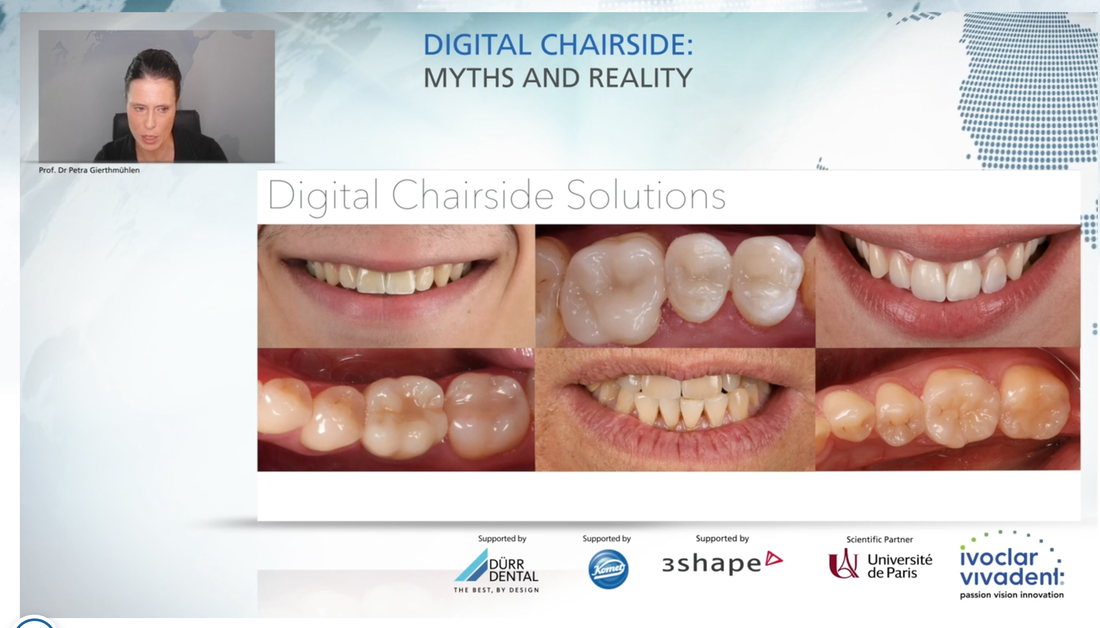
As part of Ivoclar Vivadent’s International Expert Symposium, Prof. Dr Petra Gierthmühlen gave an online presentation titled “Digital Chairside: Myths and Reality”.
By Danny Chan
The webinar provides insight into the emerging technologies for chairside dentistry – mainly that which highlights the advantages of modern CAD/CAM materials and comprehensive digital workflows.
Dr Gierthmühlen is the Professor and Chair of the Department of Prosthodontics at the Heinrich Heine University in Duesseldorf, Germany. Her lecture takes a look at the current state of chairside milling that enables faster fabrication times with protocols that are adjusted to the respective CAD/CAM material.
Based on the digital treatment concept of the Heinrich Heine University, Dr Gierthmühlen provided scientific evidence for the clinical success of CAD/CAM materials and the corresponding preparation designs.
The webinar began with a single tooth restoration case, and moved to each new case with increasing level of complexities.
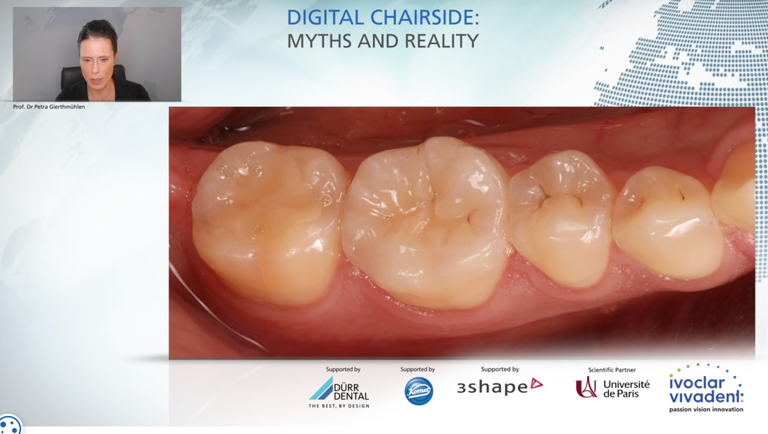
The first case, focusing on the chairside CAD/CAM enabled placement of inlays and onlays, shows the digital design of the occlusal anatomy and digital fabrication process.
Dr Gierthmühlen then provided comparison study between CAD/CAM fabricated onlays and press fabricated onlays – showing high survival rates and hardly any discernible differences in split-mouth study survival rate (100% press fabricated vs CAD/CAM fabricated 97.5%) after long-term performance (7 years).
For her next case, a Lithiumdisilicate Anterior Crown Restoration, the Professor presented on how chairside dentistry was employed to restore two fractured central incisors using a straightforward digital workflow.
Dr Gierthmühlen showed that reasonable aesthetic outcomes and high survival rates (based on long-term studies) were obtained for the endodontically-treated incisors.
The following case for Lithiumdisilicate Posterior Screw-retained Implant Restoration related to a patient who had presented with molars that needed to be extracted due to endodontical complications.
Using guided surgery along with chairside CAD/CAM dentistry, Dr Gierthmühlen showed how proper angulation of the implant and ideal screw channel axis were achieved in the digital workflow, with help from a newly acquired CEREC Omnicam.
Long-term clinical studies, based on a 7-year recall, showed the restorations remain in very good shape with no fractures at all.
The lecture also moved on to more complex cases – including that of a Lithiumdisilicate CAD/CAM Anterior implant crown.
Case: An implant that was placed to replace central incisor developed peri-implant mukositis and needed therapy to control the peri-implant infection. Once accomplished, the patient needed a new anterior crown as there was also misfit of margin within the old implant crown.
After removing the implant crown and scanning the peri-implant tissue to get an outline for the restoration, scan posts were inserted to determine location of implants. The scan posts were then transferred into software for production of zirconia abutment. At the try-in of the abutment, zirconia abutment was scanned to produce the chairside implant crown for this zirconia abutment.
The 5axis-milling machine PMOne produced a very precise crown margin.
The fit and form was excellent in blue and try-in stage followed by surface characterization, to create a beautiful final restoration.
Finally, the lecture moved on to the highest level of complexity: Full Arch cases.
Case: Patient treated more than 5 years ago presented with caries lesions; dramatically changed occlusal anatomy; dentin exposure on teeth; and suffering from hypersensitivity due to erosion.
To obtain orientation, Facescan was used to capture digital orientation of patient’s upper/ lower jaw, then on position of teeth to get full orientation of new vertical dimension of occlusion to design reference for tooth shade.
Digital mock-up and try-in was also used to develop a digitally milled prototype. This high-predictability, 100% digitally simulated workflow lets the patient decide whether he wants to proceed with treatment, based on the digital results.
The digital lab-side CAD/CAM workflow – because chairside CAD/CAM was not available at the time – was able to restore occlusal anatomy and function for patient in both upper and lower jaw and increase his vertical dimension of occlusion.
The 5-year clinical result shows high predictability for the complex full arch case: there were no fractures, gingival recession, or debonding in lower jaw. The patient also retains a similar smile line.
This is an excerpt of a webinar presented at the International Expert Symposium (IES). The IES is a digital platform Ivoclar Vivadent launched to host a series of free live-steamed and on-demand webinars.
For more information, visit: www.ivoclarvivadentacademy.com
By Danny Chan
The webinar provides insight into the emerging technologies for chairside dentistry – mainly that which highlights the advantages of modern CAD/CAM materials and comprehensive digital workflows.
Dr Gierthmühlen is the Professor and Chair of the Department of Prosthodontics at the Heinrich Heine University in Duesseldorf, Germany. Her lecture takes a look at the current state of chairside milling that enables faster fabrication times with protocols that are adjusted to the respective CAD/CAM material.
Based on the digital treatment concept of the Heinrich Heine University, Dr Gierthmühlen provided scientific evidence for the clinical success of CAD/CAM materials and the corresponding preparation designs.
The webinar began with a single tooth restoration case, and moved to each new case with increasing level of complexities.
The first case, focusing on the chairside CAD/CAM enabled placement of inlays and onlays, shows the digital design of the occlusal anatomy and digital fabrication process.
Dr Gierthmühlen then provided comparison study between CAD/CAM fabricated onlays and press fabricated onlays – showing high survival rates and hardly any discernible differences in split-mouth study survival rate (100% press fabricated vs CAD/CAM fabricated 97.5%) after long-term performance (7 years).
For her next case, a Lithiumdisilicate Anterior Crown Restoration, the Professor presented on how chairside dentistry was employed to restore two fractured central incisors using a straightforward digital workflow.
Dr Gierthmühlen showed that reasonable aesthetic outcomes and high survival rates (based on long-term studies) were obtained for the endodontically-treated incisors.
The following case for Lithiumdisilicate Posterior Screw-retained Implant Restoration related to a patient who had presented with molars that needed to be extracted due to endodontical complications.
Using guided surgery along with chairside CAD/CAM dentistry, Dr Gierthmühlen showed how proper angulation of the implant and ideal screw channel axis were achieved in the digital workflow, with help from a newly acquired CEREC Omnicam.
Long-term clinical studies, based on a 7-year recall, showed the restorations remain in very good shape with no fractures at all.
The lecture also moved on to more complex cases – including that of a Lithiumdisilicate CAD/CAM Anterior implant crown.
Case: An implant that was placed to replace central incisor developed peri-implant mukositis and needed therapy to control the peri-implant infection. Once accomplished, the patient needed a new anterior crown as there was also misfit of margin within the old implant crown.
After removing the implant crown and scanning the peri-implant tissue to get an outline for the restoration, scan posts were inserted to determine location of implants. The scan posts were then transferred into software for production of zirconia abutment. At the try-in of the abutment, zirconia abutment was scanned to produce the chairside implant crown for this zirconia abutment.
The 5axis-milling machine PMOne produced a very precise crown margin.
The fit and form was excellent in blue and try-in stage followed by surface characterization, to create a beautiful final restoration.
Finally, the lecture moved on to the highest level of complexity: Full Arch cases.
Case: Patient treated more than 5 years ago presented with caries lesions; dramatically changed occlusal anatomy; dentin exposure on teeth; and suffering from hypersensitivity due to erosion.
To obtain orientation, Facescan was used to capture digital orientation of patient’s upper/ lower jaw, then on position of teeth to get full orientation of new vertical dimension of occlusion to design reference for tooth shade.
Digital mock-up and try-in was also used to develop a digitally milled prototype. This high-predictability, 100% digitally simulated workflow lets the patient decide whether he wants to proceed with treatment, based on the digital results.
The digital lab-side CAD/CAM workflow – because chairside CAD/CAM was not available at the time – was able to restore occlusal anatomy and function for patient in both upper and lower jaw and increase his vertical dimension of occlusion.
The 5-year clinical result shows high predictability for the complex full arch case: there were no fractures, gingival recession, or debonding in lower jaw. The patient also retains a similar smile line.
This is an excerpt of a webinar presented at the International Expert Symposium (IES). The IES is a digital platform Ivoclar Vivadent launched to host a series of free live-steamed and on-demand webinars.
For more information, visit: www.ivoclarvivadentacademy.com

 RSS Feed
