By Danny Chan
Since its inception in 1916, Japanese equipment manufacturer Morita has not failed to capture our imagination with one groundbreaking innovation after another. In 1927, the company manufactured Unit A, known as the first dental treatment unit ever made in Japan. In 1964, it developed the revolutionary Spaceline, credited as the world’s first dental unit that allows patients to lie on their backs while dentists remain seated during treatment. In 1991, Morita gave us Root ZX, an epoch-making root canal measurement device.
Morita’s astounding ability to adapt in a fast-changing, technology-driven industry finds its roots in time-tested R&D philosophy, where an utter devotion to quality in product design and development trumps eagerness to flood the marketplace with standard hardware.
With the introduction of Veraviewepocs in 1997, Morita solidified its position in the dental diagnostic equipment market. Although the company’s entry into this sector began as early as 1967 with the world-renowned “Panex-E” — Morita’s first panoramic X-ray device designed for imaging the entire jawbone region while emitting extremely low radiation — the advent of Veraviewepocs marked the manufacturer’s growing confidence in offering an X-ray unit at the bleeding edge of turnkey imaging technology.
The Veraviewepocs brand also represents evolving technology; first unveiling its digital version in 1998 incorporating a PC intra-clinic LAN system (and the “Dixel” LAN systemfor digital dental X-ray images), then in 2001, adding notches to its digital capacity with caphalometric function in a high-speed (6 seconds) scanning model.
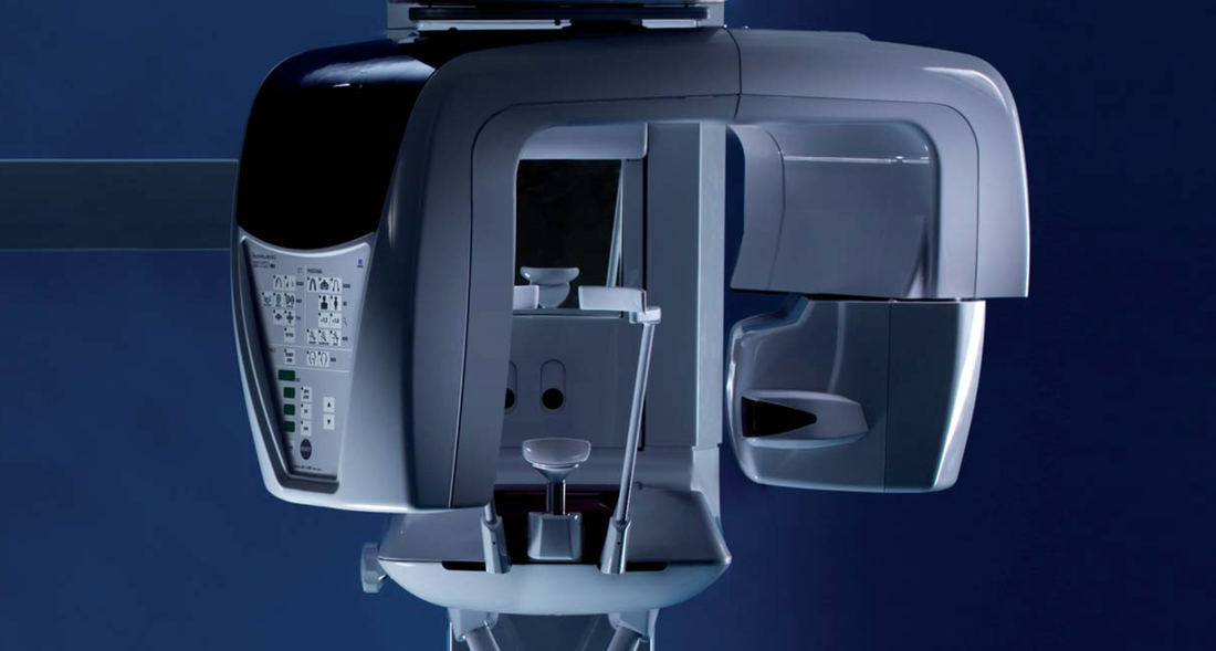
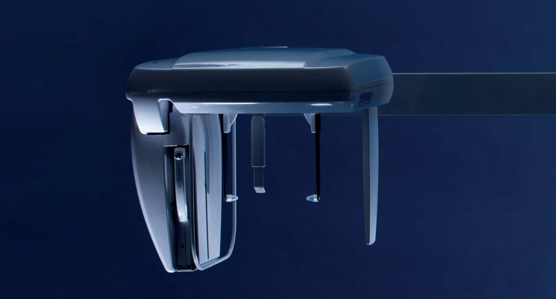
Continuing this evolution into the 21st Century is Veraviewepocs 3D, a cost-effective model that advances class-leading clarity ideal for endodontics, general dentistry and periodontics. Offering digital 3D, panoramic, and cephalometric imaging options — no cassette change required — this model features built-in sensors for all image types, designed to save time and protect the hardware. Providing Ø 40 x H 40 mm and Ø 40 x H 80 mm 3D fields of view, 3De is suitable for over 90% of all clinical cases requiring 3D analysis. In addition, it offers a ‘true’ panoramic image unlike other devices that digitally reconstruct a 3D image.
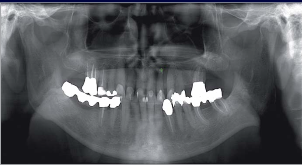
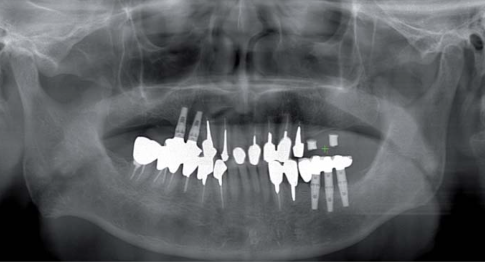
Users choose from a variety of imaging options via interchangeable cassettes. The multi-function cassette feature enables both panoramic and 3D images to be taken without changing the cassette. Contrast-rich images of both hard and soft tissue, with minimal artifacts and zero distortion, permits a highly detailed assessment — images are captured in super-high resolution (125 μm voxel). The future-proof product allows upgrade from a 2D to 3D model at a later date.
Veraviewepocs 3D does all the work
For Dr Matthew Lee, principal dentist at Sydney-based Worldciti Dental, the Veraviewepocs 3D is simply “an excellent device that makes life easier”. Having spent 17 years in private practice, Lee keenly understands the benefits of taking patient X-rays in-house on a multi-purpose unit that offers 3D images of region, panoramic and cephalometric views.
“Besides the convenience factor, I think one advantage of having an X-ray machine in the surgery is the ability to obtain the appropriate type of images for each individual case in a speedy and efficient manner,” Lee says.
“While the quality of images allows for accurate assessment, the 3D imaging capabilities virtually eliminates guesswork.”
Lee says the Veraviewepocs compares favorably with both the Sirona digital OPG, lateral Ceph and i-CAT units that he used in the past. “Having used the Veraviewepocs for 9 months, I can qualify that the images it delivers are of higher quality, and offers more accurate diagnosis.”
As with any high-end X-ray unit, superb image accuracy and fidelity, easy positioning and image-manipulation as well as versatile image processing are key components around which current imaging technology revolves. The Veraviewepocs 3D brings to bear these attributes in an intelligent package.
The integrated sensor for 3D Images and panoramic radiographs enables effortless positioning by panoramic graphic display. Before taking a 3D image exposure, the instrument releases a high resolution panoramic exposure to target the region of interest on the PC monitor.The C-arm then automatically moves into the optimum patient position to get a 3D image centered on the region of interest. This process produces images with a high degree of reproducibility.
The Veraviewepocs offers three options for accurate 3D positioning: Bi-directional scout, 5 positioning laser beams, or Morita’s unique automatic re-positioning using the high resolution panoramic as a scout image.
“Taking a 3D image is as simple as clicking on the desired position on the panoramic view.”
Lee beams: “The unit automatically adjusts to get the right exposure for the 3D image.”
“Absolute must” for implant treatment
Using the X-ray equipment mainly for implant, surgical extraction and orthodontic cases, Lee finds the Veraviewepocs 3De particularly suited for endodontic cases in revealing a problemetic root or canal. “It is also helpful in assessing the size and position of the abscess and abnormality.”
“As for implant procedures,” he emphasizes, “I believe that 3D imaging is an absolute must. For example, the Ø 40 x H 80 mm images are useful in determining the relationship of opposing teeth for dental implant planning.”
The fully digital system, says Dr. Lee, also simplifies image processing for 3D images with intelligent volume rendering and real-time reslice
“It helps that you are able to view 3D images on any computer or export them to third-party software for more specialized processing.”
Newly improved features include: Dose reduction, and a high-definition (HD) cephalometric update. Veraviewepocs 3D, and all of Morita’s 3D units, now come automatically equipped with a new dose reduction feature for increased patient protection. Across all 3D fields of view, dosage has been reduced 30% to 40%. By optimising the intensity of the X-rays, 3D is able to greatly decrease the overall level of emissions.
Morita’s latest HD update has enhanced the cephalometric image quality. The result is high definition, cephalometric images with amazing clarity and soft tissue display. This new feature, also available in Veraviewepocs 2D, makes it easier to plot key points for cephalometric measurements.
With a price point under A$90,000 and considering its marquee label standing, Morita’s 3D imaging solutions are certainly poised for the competition. As most customers would readily testify, purchase consideration for a Morita product often boils down to the company’s venerated brandname. Of course, a smaller price tag doesn’t hurt either.
Asked why he picked the Veraviewepocs 3D, Dr Lee typifies: “I simply trust Morita as a reliable brand that delivers excellent quality. It’s also a great investment.”


 RSS Feed
